

Our ability to image the subatomic realm is limited not only by resolution but also by speed. The particles that make up atoms – and that fly freely from them – can theoretically move at nearly the speed of light.
In practice, they often move much slower, but even these slower speeds are far too fast for our eyes or technology to perceive. This made observing the behavior of electrons a challenge – but now the development of a new microscope imaging technique is allowing scientists to capture them in motion in real time.
It is the work of a team of physicists at the University of Arizona Tucson led by Dandan Hui and Husain Alqattan and can capture images at the speed of attoseconds; that’s a trillionth of a second. They have called the technique attomicroscopy.
“Improving the temporal resolution in electron microscopes has been long awaited and is the focus of many research groups because we all want to see the electron motion,” says physicist Mohammed Hassan of the University of Arizona Tucson.
“These movements occur in attoseconds. But now, with our electron transmission microscope – we call it ‘attomicroscopy’ – we can achieve a temporal resolution in the attosecond range for the first time. For the first time, we can see parts of the electron in motion.”
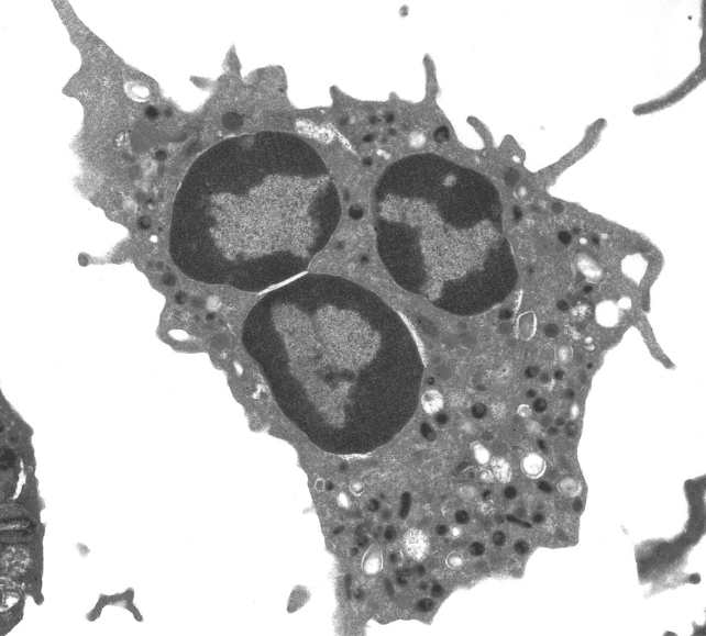
Transmission electron microscopy (TEM) is a technique used to create images of the smallest structures in the physical world. It uses electrons, not light, to create images. A beam of electrons is passed through a sample of material. The interaction between the electrons and the sample creates the image. For example, below is a TEM image of a white blood cell.
Instead of the shutter speed of a traditional camera, TEM is based on the speed of the laser pulses used to transfer the electrons. The shorter the duration of the laser pulses, the better the resulting image. So if you want better image quality, you can achieve this by developing a laser that can emit shorter pulses.
Previously, TEM lasers reached a duration of a few attoseconds, released in a series, similar to a short static burst.
This is an absolutely remarkable, Nobel Prize-worthy achievement. The problem is that although it produces a series of images, the electrons are moving slightly faster, so the changes in an electron between pulses are lost.
The researchers wanted to see if they could shorten the duration of the pulsed beam to just one attosecond—the speed at which the electrons move in the beam—so that the TEM could capture them in a still image.
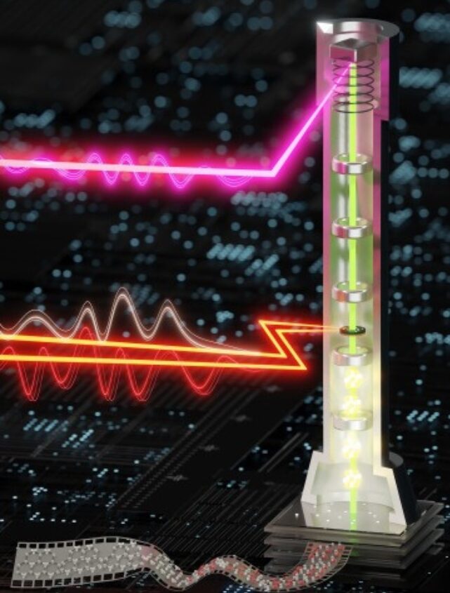
The breakthrough was achieved by splitting the pulse into three parts: two light pulses and one electron pulse. The first light pulse is called the pump pulse. It injects energy into a graphene sample, causing the electrons to jump around.
This is followed by the second pulse of light, the gate pulse, which creates a gate or window. While it is “open”, a single attosecond electron pulse is fired at the sample and the subatomic processes at attosecond speeds are recorded.
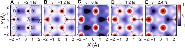
The result is a precise map of electron dynamics – a map that opens the door to new studies of the behavior of these important particles.
“This transmission electron microscope is like a very powerful camera in the latest version of smartphones. It allows us to take pictures of things we couldn’t see before – like electrons,” says Hassan.
“We hope that this microscope will enable the scientific community to understand the quantum physics behind the behavior and motion of an electron.”
The study was published in Scientific advances.
